Epileptic Disorders
MENUSeizure-induced reversible MRI abnormalities in patients with single seizures: a systematic review Volume 23, numéro 4, August 2021

- Mots-clés : seizure-induced MRI abnormality, MRI, seizure, reversible
- DOI : 10.1684/epd.2021.1300
- Page(s) : 552-62
- Année de parution : 2021
Objective
Differentiating seizure-induced reversible MRI abnormalities from MRI changes secondary to underlying cerebral pathologies can be challenging for clinicians in the investigation of seizures. The aim of this study was to delineate the characteristic features of reversible seizure-induced MRI abnormalities.
Methods
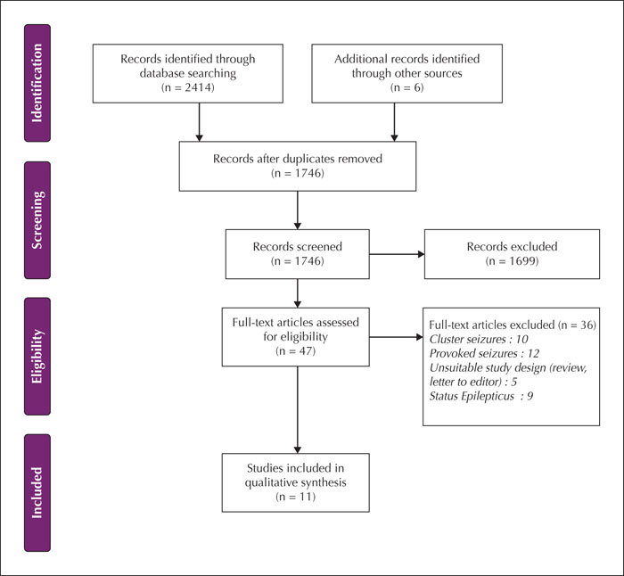
A systematic search of the databases Medline (1946-2020), PubMed (1996-2020), and Embase (1947-2020) was performed in keeping with the Preferred Items Reporting for Systematic Reviews and Meta-Analyses (PRISMA) guidelines. All publications in English, including case reports, of single unprovoked seizure patients with seizure-induced MRI abnormalities demonstrating complete resolution, were included. Two authors extracted data using a predefined template and evaluated the quality of the included studies. MRI data were additionally reviewed by a neuroradiologist. All data were synthesised qualitatively.
Results
There were 11 publications altogether, yielding a total of 27 cases that were pertinent to our research question. Abnormalities were most commonly observed on T2-weighted sequences. The most commonly observed constellations of MRI features (“composite pattern”) included the following: cortical or subcortical signal change with or without leptomeningeal enhancement, signal abnormality in the splenium of the corpus callosum, and hippocampal signal abnormality. Seizure-induced reversible MRI abnormalities were observed as early as six hours from seizure onset and resolved completely as early as five days from seizure onset. A key limitation of this systematic review was the variability and incomplete reporting of clinical data, especially with regards to seizure semiology and MRI sequences performed, which may have limited our ability to make more definitive conclusions.
Conclusions
Seizure-induced reversible MRI changes may appear within hours of seizure onset and resolve within a variable time frame, ranging from days to weeks. Bilateral seizure-induced reversible MRI abnormalities tend to be associated with generalised seizures while unilateral seizure-induced reversible MRI abnormalities may be associated with focal seizures, usually ipsilateral to the seizure focus.