Epileptic Disorders
MENUDefinition of the neurological phenotype associated with dup (X)(p11.22-p11.23) Volume 13, numéro 3, Septembre 2011

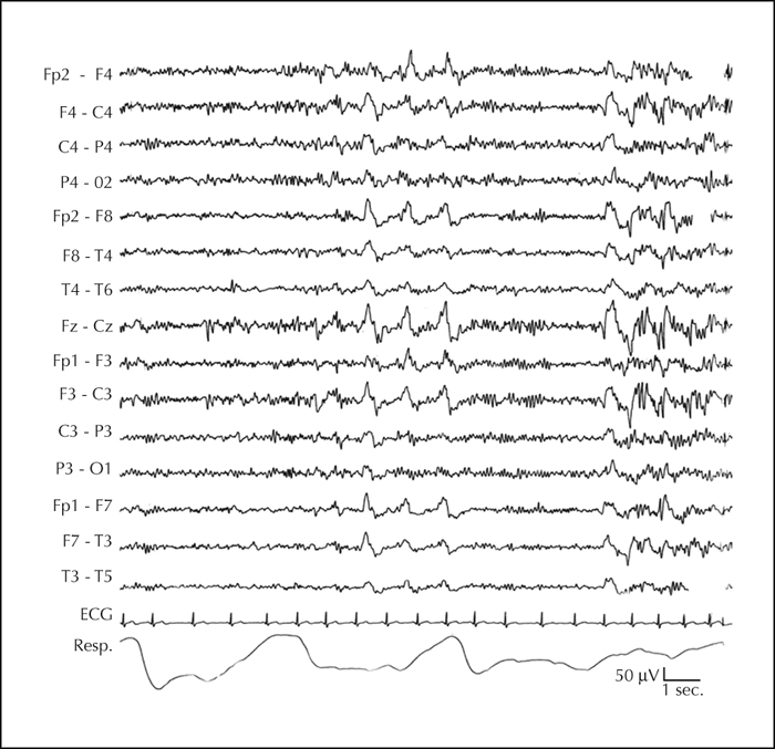
Figure 1 EEG of Case II.1 (family 2) during sleep deprivation. During sleep (up to phase 2 NREM), high amplitude bisynchronous anterior dominant spike-wave complexes are evident.
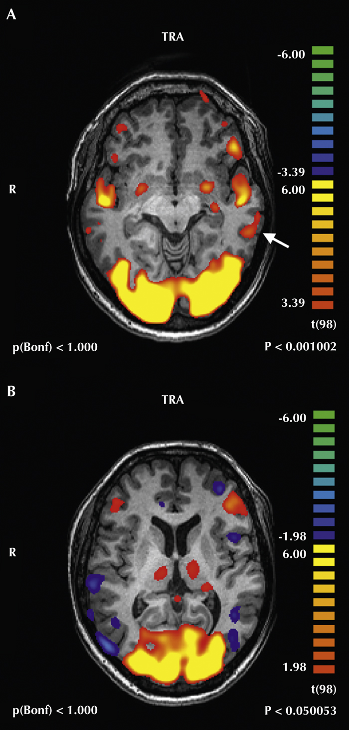
Figure 2 fMRI of Case II.1 (family 2); axial representation of the eloquent areas was evoked by two language paradigms. A) Text listening (p<0.001). Besides the wide occipital bilateral activations, bilateral temporal activations are appreciable; on the left side, an additional posterolateral eloquent area (arrow) is evident. B) Image naming (p<0.05). Wide bilateral occipital activations are appreciable together with smaller frontal opercular eloquent areas prevailing on the left side. Note the threshold had to be lowered (p<0.05 vs p<0.001 in A) to disclose eloquent areas. Asymmetries can be seen in the language eloquent areas indicating a left hemispheric dominance and a smaller width of the language eloquent areas.
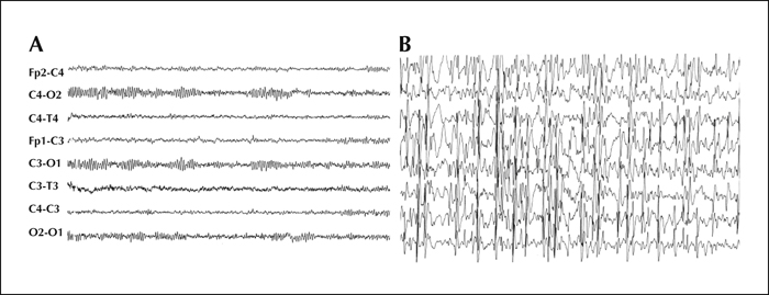
Figure 3 EEG of sporadic Case 1. During the awake state (A) specific discharges were not present, whereas during sleep (B) generalised epileptiform discharges became subcontinuous and ESES was determined.
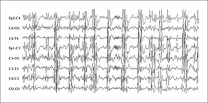
Figure 4 EEG of sporadic Case 1. As in figure 3, the EEG recording shows the persistence of sporadic spindles.
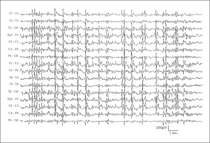
Figure 5 EEG of sporadic Case 2. During drowsiness and slow sleep, generalised epileptiform discharges, such as polyspikes and slow waves, became subcontinuous and ESES was determined.