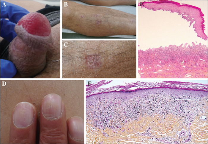
Physical examination reveals a large, painless erythematous erosion of the glans penis (A), non-specific, slightly atrophic lesions of the anterior shins, with presence of milia (B, C), and nails with grooves and ridges (D). E) Histopathological examination of the biopsy from the glans penis reveals a subepidermal blister, with an overlying thick parakeratotic epithelium and chorium containing an inflammatory infiltrate composed of lymphocytes, plasma cells and eosinophils. F) Histopathological examination of the biopsy taken from the shin reveals a moderately-thickened epidermis with an overlying orthokeratotic horny layer, vacuolated basal cell layer, and upper dermis containing a dense, band-like inflammatory lymphocytic infiltrate hugging the basal epidermal layer.