Annales de Biologie Clinique
MENUProtoscolex d’Echinococcus granulosus chez un patient atteint de kyste hydatique pulmonaire Volume 78, numéro 4, Juillet-Août 2020

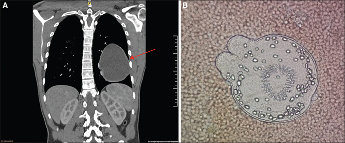
Figure 1
(A) Computed tomography of the chest with the hydatid cyst in the left lung (arrow). (B) Echinococcus granulosus protoscolex in the cystic content observed under the microscope at 400x magnification (surrounded by many red blood cells).

