Epileptic Disorders
MENUVisual word form area hyperexcitability associated with focal epileptiform activity in a case of reading epilepsy Volume 24, numéro 6, December 2022

- Mots-clés : reading epilepsy, reflex epilepsy, visual word form area, functional connectivity, adaptative direct transfer function
- DOI : 10.1684/epd.2022.1496
- Page(s) : 1095-101
- Année de parution : 2022
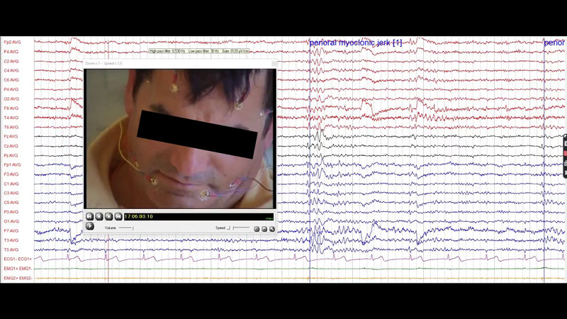
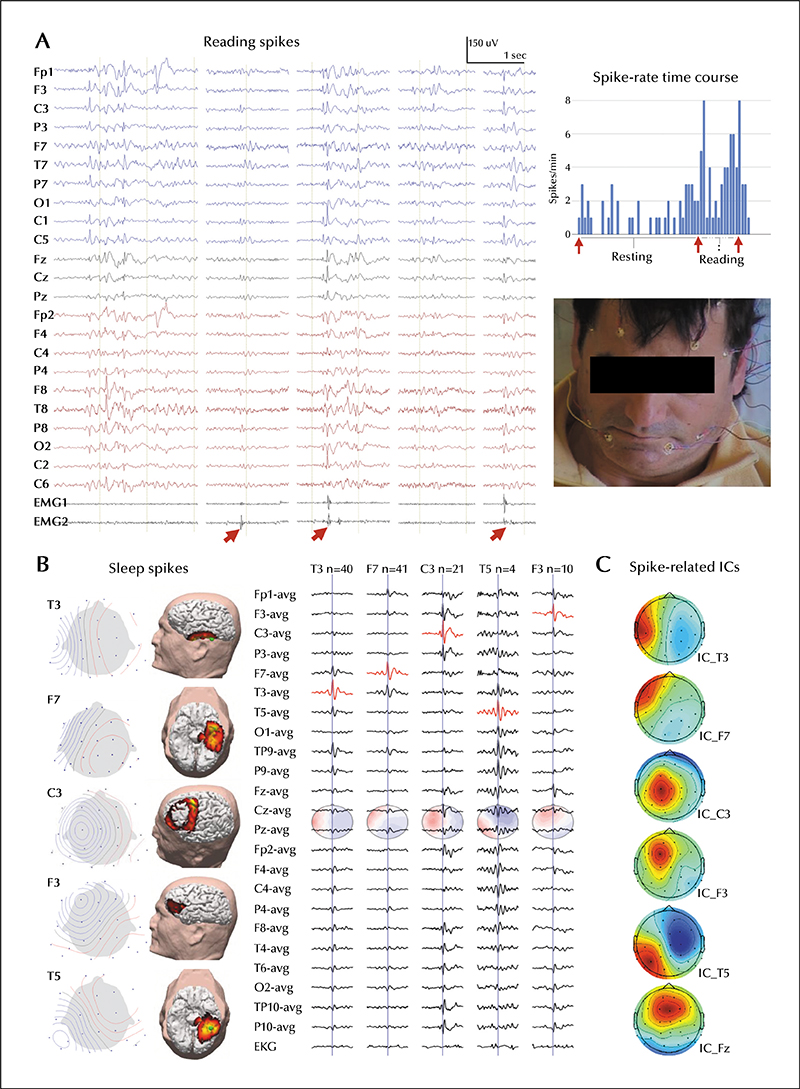
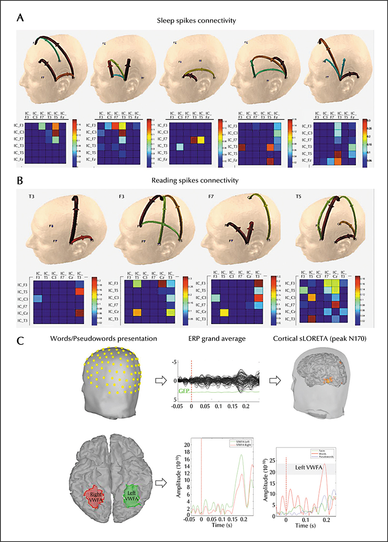
Reading epilepsy recruits critical language-related areas, with synchronization and subsequent spreading of excitation in response to the epileptogenic stimulus. The mechanism by which possible generalized discharges result in the expression of bilateral or unilateral clinical symptoms remains controversial. The cortical and subcortical areas involved may constitute part of the normal reading network, such as the visual word form area (VWFA). A right-handed, 59-year-old man was diagnosed with epilepsy at the age of 15 after tonic-clonic seizures. Later, the patient described myoclonic jerks of the masticatory and perioral muscles while reading. A multimodal approach with magnetic resonance imaging and ambulatory and video-electroencephalogram was used for seizure characterization and source analysis. A left hemisphere spontaneous occipitaltemporal epileptic focus, activated by reading, was observed, spreading broadly throughout frontal and temporal language networks. There was an abnormally increased cortical response to visual word presentation in comparison to pseudowords. Spatial localization of spike sources suggested a close association between the primary epileptic focus and the VWFA. This epileptiform activity seems to be selectively triggered at an early stage of lexical processing, with a functional connection between the epileptic network and the VWFA. This multimodal and functional connectivity approach could be helpful in determining the epileptic network in reading epilepsy.