Epileptic Disorders
MENUReflex operculoinsular seizures Volume 18, numéro 1, March 2016

Figure 1
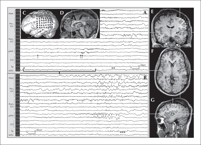
Invasive EEG recording of somatosensory evoked seizure in Patient 1. The recording sites are shown on a 3-D representation of the patient's brain in insets (C) and (D). In the upper EEG recording, rubbing the patient's right hand (*indicates onset of stimulus; horizontal bracket shows the duration of stimulation) triggers low-amplitude preictal spikes (↑) in the left posterior long insular gyrus (triangulated in the insets: [E] coronal view, [F] axial view, and [G] sagittal view) and frontal operculum, transitioning into a low-voltage fast activity ictal pattern (↑↑). Clinically, the patient feels right hemibody pain and lifts his right arm (**) until the end of the ictal discharge (***lower EEG recording). The latency between the trigger and the first epileptiform discharge was 2.850 seconds. PMA: premotor area; Pre-CG: precentral gyrus; Post-CG: postcentral gyrus; pINS: posterior insula; CG: cingulate gyrus; Tp: temporal pole; MTG: middle temporal gyrus.

Figure 2
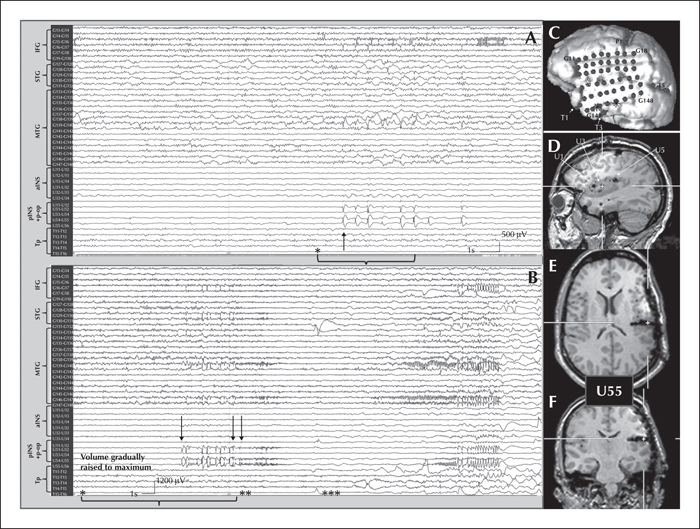
Invasive EEG recording of reflex audiogenic operculoinsular spikes (A) and seizure (B) in Patient 2. The recording sites are shown on a 3-D representation of the patient's brain in inset (C). In the upper EEG recording (A), an auditory stimulus to the left ear (2000 Hz, 85 dB) (*indicates onset of auditory stimulation; horizontal bracket indicates duration of stimulus) triggers, within a second, asymptomatic rhythmic spike-and-wave discharges (↑), maximum over the left posterior insula (as triangulated in the insets: [D] sagittal view, [E] axial view, and [F] coronal view) and parietal more than temporal opercula. High-pass filter: 0; low-pass filter: 35 Hz. In the lower EEG recording (B), listening to a piece of classical music gradually raised to maximum volume (*indicates onset; horizontal bracket shows stimulus duration) triggers a similar burst of spikes (↑) transiting into a seizure (↑↑) with the appearance of a high-frequency discharge in the posterior insula, parietal and temporal opercula. Clinically, the patient suddenly feels his aura, quickly removes his headphones (**), but is unable to abort subsequent evolution into complex motor behaviours (***). High-pass filter: 0 and low-pass filter: 20. IFG: inferior frontal gyrus; STG: superior temporal gyrus; MTG: middle temporal gyrus; aINS: anterior insula; pINS: posterior insula; P-op: parietal operculum; Tp: temporal pole.

