Epileptic Disorders
MENUIctal dysprosody and the role of the non-dominant frontal operculum Volume 7, numéro 3, September 2005

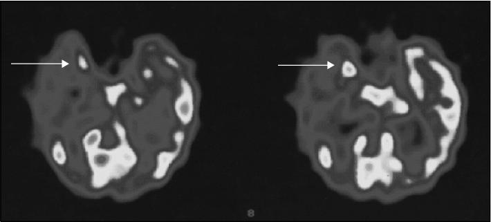
Figure 1 Ictal SPECT (Neurolithe 99mTc) showed an increased blood flow in the right mesial temporal lobe.
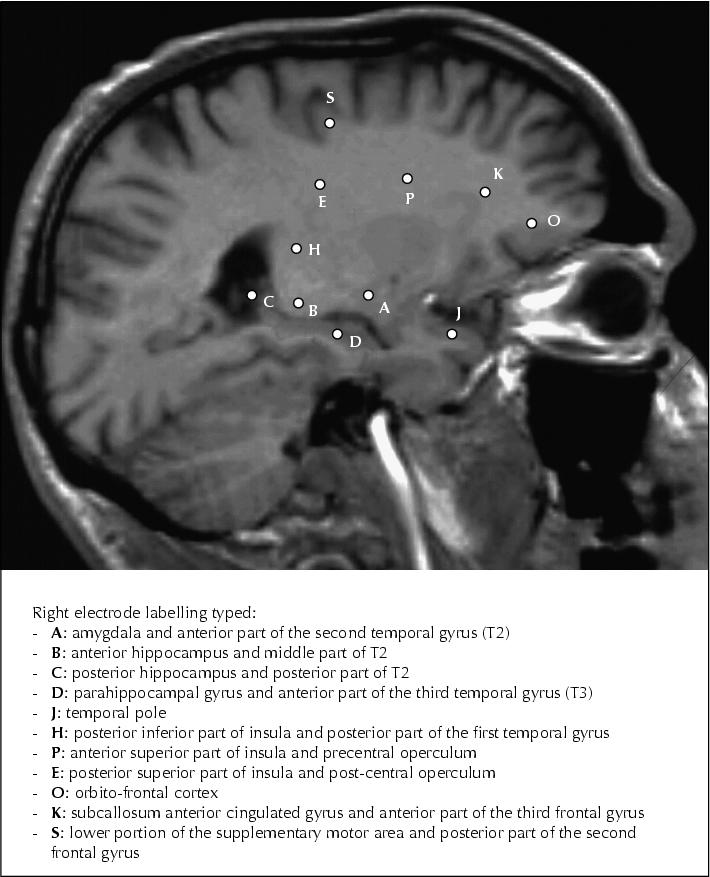
Figure 2 Eleven electrodes were placed in the right hemisphere and one electrode was placed in the left amygdala.
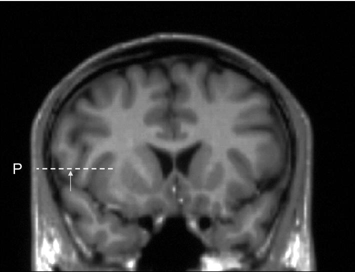
Figure 3 Precentral operculum electrode: ictal recurrent speech utterances associated with an altered prosody occurred during both spontaneous seizures and a very localized stimulation-induced discharge of the non-dominant precentral operculum.