Epileptic Disorders
MENUGood outcome in adult-onset Rasmussen's encephalitis syndrome: is recovery possible? Volume 17, numéro 2, June 2015

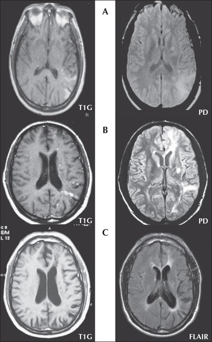
Figure 1
(A) Prodromal phase; MRI showing cortical swelling and hyperintense areas in the left temporal and parietal lobes. Contrast enhancement is clear in the left image. (B) Prodromal/acute phase transition; MRI showing cortical-subcortical hyperintense lesions in the left hemisphere. Contrast enhancement is clear in the left image. An initial atrophy of the left hemisphere is also present. (C) Residual phase; MRI showing left hemispheric atrophy with no contrast enhancement. Atrophy of the head of the left caudate nucleus is also evident. PD: proton density; T1G: T1-weighted gadolinium MRI; FLAIR: fluid-attenuated inversion recovery.

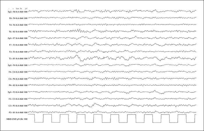
Figure 2
EEG showing slow activity confined to the left hemisphere.

