Epileptic Disorders
MENUExtraoccipital photoparoxysmal response in a case of focal encephalitis Volume 13, numéro 1, Mars 2011

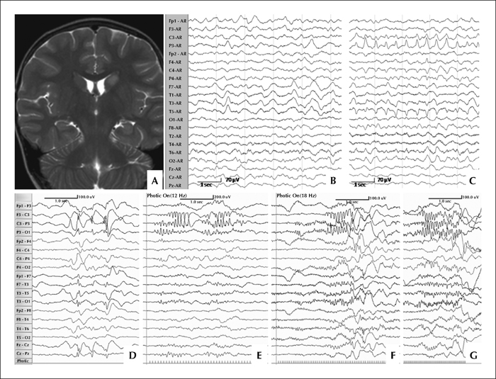
Figure 1 A) Coronal T2 MRI showing left caudate hyperintensity during acute phase of the illness. B) Initial EEG showing left parieto-temporal slowing (B) and (C) left parietal PLEDs. D-G) EEG at six months follow-up showing (D) left centro-parietal spike-and-wave discharges and focal photoparoxysmal response at the left centro-parietal region with 12âHz (E), 18âHz (F) and 30âHz (G) photic frequencies. See text for further details.