Epileptic Disorders
MENUAsymmetric tonic seizures with bilateral parietal lesions resembling frontal lobe epilepsy Volume 3, numéro 1, Mars 2001

Patient
Patient 1 was a 24-year-old, right-handed man with intractable seizures since the age of 14 years. The patient was the product of a difficult labor; he suffered neonatal hypoxia, and convulsions occurred until several months after delivery. Since the age of 14 years, generalized tonic-clonic seizures occurred occasionally. At age 18, the patient started having the current seizure type. Initially, a sharp lightning sensation occurred in the whole mouth and in the pharyngeal area, immediately followed by bilateral asymmetric hand abduction, thrashing movements of both feet, and speech arrest. The episode lasted for less than a half minute, and he was completely aware throughout the episode. The patient had been treated with phenobarbital 150 mg/day (trough blood level of 34.3 mg/L), carbamazepine 400 mg/day (2.5 mg/L), valproic acid 1000 mg/day (68.9 mg/L) and phenytoin 200 mg/day (9.7 mg/L) in various combinations. However, his seizures occurred several times a day, with the maximum frequency of 10 times a day. Neurologically, moderate mental retardation (total IQ of 42, verbal IQ of 49 and performance IQ less than 45 in the Wechsler Adult Intelligence Scale-Revised: WAIS-R), mild dysarthria, mild gait ataxia and mild postural and action tremor of hands were observed. Interictal EEG showed slow, disorganized posterior dominant rhythm (7-8 Hz), and diffuse, intermittent, irregular slow activity, more on the right hemisphere, but no apparent epileptiform discharges.
Patient 2 was a 32-year-old, right-handed man with intractable seizures since the age of 7 years. The patient was a low birth weight infant born prematurely. No febrile convulsions occurred. The patient had generalized convulsions with high fever at the age of 7 years, and then his school performance became poor. Since the age of 18, the current seizure type started. Ictal symptoms started with a brief, sharp, lightning sensations in the whole body, followed by asymmetric tonic hand posturing (bilateral hand abduction, left elbow flexion, right elbow extension), head and eye deviation to the right, thrashing movements of both feet and inability to speak. The episode lasted for less than a half minute, and about 70% of the time he was aware throughout the episode. About two thirds of the time, seizures were apparently provoked by unexpected stimuli such as a loud noise, a call or tapping his shoulder from the back. No seizures were induced by visual stimuli. The patient was treated with carbamazepine 800 mg/day (trough blood level of 7.4 mg/L) and zonisamide 600 mg/day (11.6 mg/L) on admission, and had previously received phenytoin 350 mg/day, phenobarbital 50 mg/day, carbamazepine 600 mg/day, clonazepam 3 mg/day and valproic acid 600 mg/day in various combinations. Nevertheless, his seizures have been poorly controlled, with an average frequency of 2-3 times a day, the maximum frequency being 10 times a day, and the longest seizure-free interval being 3 days. The patient also had generalized tonic-clonic seizures immediately following the above seizure type, several times per year. Neurologically, moderate mental retardation (total IQ of 56, verbal IQ of 59 and performance IQ of 59 in WAIS-R), leftward gaze-evoked horizontal nystagmus, homonymous right inferior quadrantapnosia and right eye nasal anopsia, mild dysarthria, and mild gait ataxia were observed. Interictal EEG showed slow, poorly organized posterior dominant rhythm (7 Hz), diffuse intermittent irregular slow activity, and rare, focal epileptiform discharges in the left frontal area.
Long-term video/EEG monitoring and imaging studies
A long-term video/ EEG monitoring was carried out for a total of 5 days in a hospital room with shallow cup electrodes attached by collodion in accordance with the International 10-20 System. In Patient 2, EEGs were recorded on magneto-optical disks using digital EEG equipment, for further analysis with montage reformatting and filter change. Cranial MRI and interictal single photon emission computed tomography (SPECT) (Tc-HMPAO in Patient 1 and Tc-ECD in Patient 2) were investigated. In Patient 2, ictal SPECT by means of Tc-ECD was recorded when spontaneous habitual seizures occurred. 18F-Fluorodeoxyglucose positron emission tomography (FDG-PET) and flumazenil-PET were also performed in Patient 2 to delineate an epileptogenic zone.
Ictal semiology and ictal EEG
In both patients, asymmetric tonic seizures were documented simultaneously by both EEG and video. As described in the clinical history, common to both patients was a lightning sensation in the whole mouth or in the whole body preceded asymmetric tonic posturing of the hands and thrashing movements of the feet. Usually, no apparent loss of awareness was observed, and seizures lasted for less than half a minute. No nocturnal predominance in seizure occurrence was present during the monitoring, and the seizures tended to occur in clusters in the morning in both patients.
On EEG, in Patient 1, about 18 sec after the clinical onset, relatively low amplitude, rhythmic 6-7 Hz activity was observed mainly in the parasagittal central region (figure 1). In Patient 2, more than 10 habitual seizures were recorded by a digital EEG, and no apparent ictal EEG patterns were observed even by changing montage (employing bipolar transverse or midline-longitudinal derivations) and filter setting. Only a diffusely attenuated EEG background in association with significant EMG and movement artifacts were observed throughout the ictal period. One seizure was provoked by an unexpected sound which occurred when his mother put a coffee cup on the table, and neither clear transient activity in association with this sound nor the following focal ictal pattern was recognized.
Neuroimaging studies
A cranial MRI in Patient 1 showed high intensity signal abnormality in the gray and white matters of bilateral parieto-occipital to posterior temporal areas on the T2-weighted image, that appeared as a low signal intensity abnormality on the T1-weighted image (figure 2a). Interictal SPECT in Patient 1 showed low perfusion in both parieto-occipital areas.
In Patient 2, a cranial MRI showed a discrete, multilobed T1-low intensity abnormality surrounded by a T2-high intensity abnormality in the white matter in the bilateral parieto-occipital areas, which extended to both mesial occipital areas, more on the left side (figure 2b). Interictal SPECT revealed low perfusion in both parieto-occipital areas, more significantly on the left side (figure 3). Ictal SPECT clearly showed high uptake areas in the right parietal area, and also in the right frontopolar area to a similar degree (figure 3). Both FDG- and flumazenil-PET showed low activity in the bilateral parieto-occipital areas, but not in the frontal area (figure 4).
In these two patients, the following common characteristics in the clinical history, ictal semiology and neuroimaging findings were seen. (i) Clinically, both patients had significant perinatal brain insults which was most likely related to the etiology of their seizure disorders, and they had moderate mental retardation. (ii) Predominant ictal semiology consisted of asymmetric tonic posturing of the hands and thrashing movements of the feet, which was clinically consistent with seizures arising from the SMA. (iii) Consistently, their seizures were preceded by a bilateral lightning sensation in body parts. (iv) Neuroimaging studies suggested clear lesions in both parieto-occipital areas, but no apparent lesions in the mesial frontal or even lateral frontal areas. (v) Seizures tended to occur in clusters but not predominantly during the nocturnal period.
Based on the above common findings, we hypothesize that an epileptogenic zone in these two patients is present in both parietal areas, and that when ictal activity in the parietal area spreads to the frontal, at least probably into the mesial frontal, area, then clinical SMA seizures occur. In other words, we hypothesize that in the two patients, the epileptogenic zone is in the parietal area whereas the symptomatogenic zone is in the frontal area. With regard to the ictal semiology, their initial seizure symptom was a bilateral lightning sensation which probably reflects the involvement of either the primary or second somatosensory areas bilaterally. Although seizures arising from SMA may also have preceding sensory auras (tension, pulling sensation, heavy sensation, urge to move, tingling sensation in the extremities) [4], these seem more ambiguous or more related to motion sense, whereas the present patients commonly had a sharp, lightning sensation.
In Patient 2, ictal SPECT showed high uptake in the right parietal area whereas low uptake was seen during the interictal period. The right frontal area also showed high uptake during the ictal period, whereas interictally it showed no hypoperfusion by SPECT. FDG- and flumazenil-PET interictally showed low activity in both parietal areas, but not in the frontal area. Since both FDG- and flumazenil-PET could delineate an epileptogenic zone [5], it is most likely that the frontal area is not primarily an epileptogenic area but is secondarily activated by the primarily active parietal area. Furthermore, asymmetric tonic posturing without loss of awareness following lightning sensation was consistent with SMA seizures, and thrashing movements of both feet also reflect involvement of the SMA in the form of a hypermotor seizure [6]. However, those seizures did not occur nocturnally but rather in the morning during video/ EEG monitoring, which is unlike the seizures of frontal lobe epilepsy. All these findings support our hypothesis that in the two patients, an epileptogenic zone was in the parietal area whereas the symptomatogenic zone was in the frontal area.
In this hypothesis, the mechanism by which bilateral parietal lesions cause the ictal semiology of SMA seizures remains uncertain. Unilateral parieto-occipital lesion usually produces focal seizure disorders strongly related to parietal functions like somatosensory seizures by the anterior parietal area, or complex partial seizures by the posterior parietal area [7]. In the present patients, it is speculated that an ictal activity located in either side of the parietal lesions (right parietal lesion in Patient 2), quickly spread to the frontal area via the superior longitudinal fasciculus, therefore giving rise to the ictal semiology of SMA seizures. A similar ictal propagation via the superior longitudinal fasciculus, fronto-occipital fasciculus and inferior longitudinal fasciculus [8] may also explain the non-occipital clinical manifestations in patients with occipital lobe epilepsy. Out of 82 patients with parietal lobe epilepsy treated surgically, 22 (28%) reportedly had tonic posturing of extremities [9], but most patients had tonic posturing of the extremity contralateral to the epileptogenic side, and only 3 out of 82 patients (3.7%) had bilateral tonic posturing (patients 22, 53 and 74 in the appendix of Salanova et al. [9]). Those 2 patients had unilateral atrophy, one of which was revealed by pneumoencephalogram. No invasive recording was done in their series to prove the involvement of the SMA [9]. Previously, ictal spreading from parietal to frontal area in patients with asymmetric tonic posturing was reported following invasive recording in only 2 patients [10]. At least in Patient 2 in the present study, we could successfully discover the involvement of both the right parietal and the right frontal areas. The corpus callosum contains commissural fibers connecting the homologous areas of the two hemispheres. These exert not only an excitatory, but also an inhibitory effect as shown by suppression of the development of convulsive seizures by amygdaloid kindling and of ictal march in rolandic seizures in animal experiments [11]. Therefore, we also hypothesize that the presence of bilateral, homologous parietal lesions could disinhibit ictal activity on either side.
In Patient 2, the seizures were often provoked by unexpected external stimuli such as a loud noise or tapping from the back. A similar patient with asymmetric tonic seizures provoked by external stimuli, i.e. startle-provoked SMA seizures, was previously studied by invasive subdural recording that delineated an extensive ictal onset zone in the dorsolateral and mesial frontal areas [12]. The authors concluded that an extensive area of abnormally excitable tissue in the frontal lobe is essential for generation of startle-induced seizures. Previously, startle-induced seizures were observed in patients with severe neurological deficits and extensive cortical lesions [13]. In common with these previous reports, the present patients had moderate mental retardation and bilateral parieto-occipital lesions, although no lesions were present in the frontal lobe of our patients.
Differentiation of the symptomatogenic zone from the epileptogenic zone is clinically useful to better understand the pathophysiology of seizure disorders similar to those in the present patients [14]. Usually, the two zones are identical or very close to each other, but may be at times distant, overlying different lobes, in which case the diagnosis of seizure type and epilepsy can be easily erroneous, and only invasive recording could prove it [15]. In our patients, we could differentiate those two zones in the parietal and frontal areas, based on the clinical and physiological studies including various neuroimaging, EEG, MRI and ictal semiology.
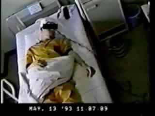
Videotape legend
The videotape includes the typical seizures (2 seizures for each patient) of both cases 1 and 2. In case 1, seizures consist of facial grimace and thrashing movements of both feet, lasting less than 1 min. In case 2, facial grimace and asymmetric tonic posturing of the extremities are followed by thrashing movements of both feet. The 2nd seizure of case 2 was evoked by an unexpected sound which occurred when a coffee cup was put on the table.
CONCLUSION
Acknowledgements:
This study was supported by Grants-in-Aid for Scientific Research (C) (2) 12210012 from the Japan Ministry of Education, Science, Sports and Culture, Research for the Future Program from the Japan Society for the Promotion of Science JSPS-RFTF97L00201.