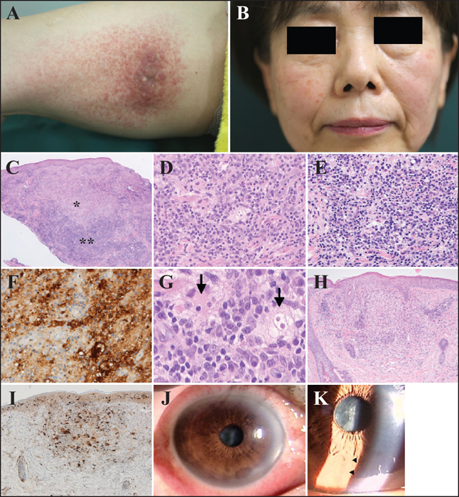
A, B) Clinical appearance of the skin lesions at the first visit. A) A rectangular patch measuring 20 cm×12 cm with a poorly defined border on the inner side of the right thigh. The rectangular patch consisted of a cluster of light violet-red macules, less than 0.5 cm in diameter, or papules. On the cranial side of the rectangular patch, there was a violet-red plaque, measuring 6.5 × 3.5 × 0.5 cm. Diffuse erythema was present on both cheeks. B) Erythematous papules were scattered on the erythema. C-G) Histopathological findings of the nodule on the right thigh. C) Two kinds of region (*, **) were observed in the dermis (haematoxylin and eosin; original magnification: ×40). D) The region indicated by * shows a dense infiltrate of large macrophages (haematoxylin and eosin; original magnification: ×400). E) The region indicated by ** shows a dense infiltrate of lymphocytes and plasma cells (haematoxylin and eosin; original magnification: ×400). F) Macrophages in (D) were positive for S100 protein (original magnification: ×400). G) Some macrophages showed emperipolesis (arrows) (original magnification: ×1,000). H, I) Histopathological findings of a papule on the right cheek. H) A nodular infiltration of macrophages, intermingled with diffuse infiltration of lymphocytes and neutrophils, was present in the superficial to middle dermis (haematoxylin and eosin; original magnification: ×40). I) Macrophages in (H) were positive for S100 protein (original magnification: ×400). J, K) Slit-lamp findings of the left eye at the first examination. Bulbar ciliary injection (J) and mutton-fat keratic precipitates (arrow heads) (K) due to anterior granulomatous uveitis were noted.
Rosai-Dorfman disease (RDD) was originally described as a rare pseudolymphomatous disorder, characterised by persistent massive lymphadenopathy and caused by increased numbers of macrophages within lymph node sinuses [1, 2]. These macrophages are often large and exhibit abnormal behaviour, taking intact cells, such as lymphocytes, erythrocytes, and plasma cells, into the cytoplasm, as a phenomenon known as emperipolesis. Population sites of these abnormal macrophages are not confined to lymph node [...]