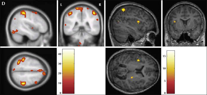
(A) Interictal SPECT showing perfusion changes (rCBF) in the bilateral frontal region, more prominent on the left than the right side (yellow arrows; coronal image), and left parietal region (yellow arrows; axial plane and sagittal plane). (B) Ictal SPECT showing perfusion changes (rCBF) in the bilateral frontal (coronal image), temporo-parietal (sagittal image), and bilateral fronto parietal regions with thalamic involvement (axial image). (C) Interictal EEG recording during simultaneous fMRI acquisition after cardio-ballastic and gradient artefact corrections. Increased delta and theta activity with spike-and-slow-wave pattern of IEDs is noted. (D) Location of peaks of regional activation using EEG-fMRI (MNI stereo tactic coordinates, p<0.001 FDR uncorrected): the left perirolandic, left post central gyrus, and bilateral parietal lobes involving the superior and inferior parietal lobule, precuneus, and right thalamus.
The common network revealed by EEG-fMRI and SPECT (A) corresponded to the left frontal parietal network.
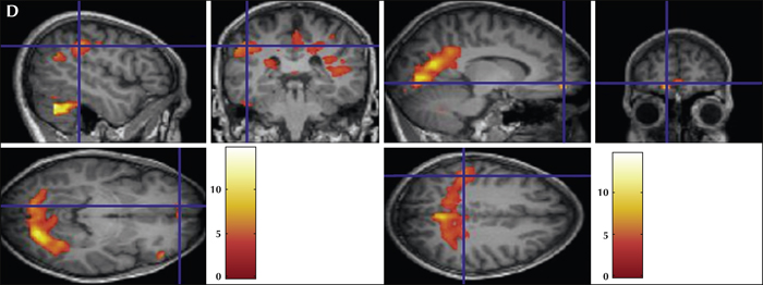
(A) Interictal SPECT showing perfusion changes (rCBF) in the left frontal, left perirolandic (yellow arrow; coronal plane), left parietal (yellow arrow; axial plane), and occipital areas (yellow arrow -sagittal plane). (B) Ictal SPECT showing perfusion changes (rCBF) in the bitemporal (coronal plane), occipital (sagittal plane), and bilateral fronto-parietal (axial plane) areas. (C) Interictal EEG recording during simultaneous fMRI acquisition after cardio-ballastic and gradient artefact corrections. Generalised sharp wave discharges are noted. (D) Location of peaks of regional activation using EEG-fMRI (MNI stereo tactic coordinates, p<0.001 FDR uncorrected.): the left fronto-temporal (coronal plane), bilateral parietal areas (coronal plane), and mesial structures, such as the paracentral lobule, cingulate, medial frontal gyrus, and lingual and medial occipital gyrus (axial and sagittal plane).
The common network revealed by EEG-fMRI and SPECT (A) corresponded to the left frontal parietal occipital network.
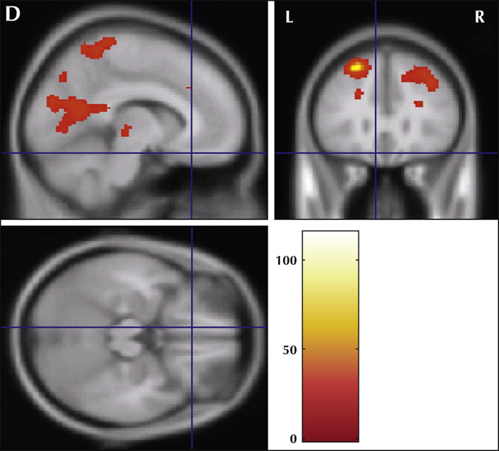
(A) Interictal SPECT showing perfusion changes (rCBF) in the bilateral frontal (yellow arrow; axial plane), biparietal (axial plane), temporal (yellow arrow; coronal plane), and occipital (sagittal plane) areas. (B) Ictal SPECT showing perfusion changes (rCBF) in the bilateral frontal, parietal, occipital (axial and sagittal plane), and bitemporal (coronal plane) regions. (C) Interictal EEG recording during simultaneous fMRI acquisition after cardio-ballastic and gradient artefact corrections. Spike-wave complex IEDs are noted. (D) Location of peaks of regional activation using EEG-fMRI (MNI stereo tactic coordinates, p<0.001 FDR uncorrected.): the left superior and inferior frontal gyrus, mesial structures (such as the paracentral lobule), cingulate gyrus, medial frontal gyrus, and lingual and medial occipital gyrus.
The common network revealed by both EEG fMRI and SPECT (A) corresponded to the fronto-parietal occipital areas.
1 Cognitive Neuroscience Center, National Institute of Mental Health, Neuro Science (NIMHANS)
2 Dept of Neuroimaging and Interventional Radiology, National Institute of Mental Health and Neuro Science (NIMHANS)
3 Dept of Neurology, National Institute of Mental Health and Neuro Science (NIMHANS), Bangalore, India
* Correspondence: Rose Dawn Bharath
Cognitive Neuroscience Center and Dept of Neuroimaging and Interventional Radiology,
National Institute of Mental Health and Neuro Science (NIMHANS),
Hosur Road,
Bangalore 560029, India
Measuring neuro-haemodynamic correlates in the brain of epilepsy patients using EEG-fMRI has opened new avenues in clinical neuroscience, as these are two complementary methods for understanding brain function. In this study, we investigated three patients with drug-resistant reflex epilepsy using EEG-fMRI. Different types of reflex epilepsy such as eating, startle myoclonus, and hot water epilepsy were included in the study. The analysis of EEG-fMRI data was based on the visual identification of interictal epileptiform discharges on scalp EEG. The convolution of onset time and duration of these epilepsy spikes was estimated, and using these condition-specific effects in a general linear model approach, we evaluated activation of fMRI. Patients with startle myoclonus epilepsy experienced epilepsy in response to sudden sound or touch, in association with increased delta and theta activity with a spike-and-slow-wave pattern of interictal epileptiform discharges on EEG and fronto-parietal network activation pattern on SPECT and EEG-fMRI. Eating epilepsy was triggered by sight or smell of food and fronto-temporal discharges were noted on video-EEG (VEEG). Similarly, fronto-temporo-parietal involvement was noted on SPECT and EEG-fMRI. Hot water epilepsy was triggered by contact with hot water either in the bath or by hand immersion, and VEEG showed fronto-parietal involvement. SPECT and EEG fMRI revealed a similar fronto-parietal-occipital involvement. From these results, we conclude that continuous EEG recording can improve the modelling of BOLD changes related to interictal epileptic activity and this can thus be used to understand the neuro-haemodynamic substrates involved in reflex epilepsy.