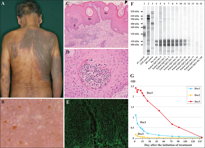
A) Clinical findings; dark-brown papillomatous vegetating plaques with pustules on the back and upper arms. B) Dermoscopy examination of the right back showing white round structures and brown, irregularly shaped structures. C, D) Histopathological findings of the right back. C) Irregular acanthosis, intraepidermal pustules, and horn cysts in the epidermis, with opening of horn cysts into the surface of the epidermis (arrowhead) (hematoxylin-eosin stain; original magnification: ×100). p: pustule; hc: horn cyst. D) Pustule filled with numerous eosinophils, neutrophils, and acantholytic keratinocytes (hematoxylin-eosin stain; original magnification: ×400). E) Direct immunofluorescence showing punctate cell surface deposition of IgG over the entire epidermis. F) Immunoblotting of normal human epidermal extracts for IgG autoantibodies. Pemphigus vulgaris (PV) control serum reacts with 160-kDa Dsg1 and 130-kDa Dsg3 (lane 1). Paraneoplastic pemphigus (PNP) control serum reacts with 210-kDa envoplakin and 190-kDa periplakin (lane 2). Bullous pemphigoid (BP) control serum reacts with 230-kDa BP230 and 180-kDa BP180 (lane 3). Anti-Dsc monoclonal antibody reacts with the 110-kDa a-form and the 100-kDa b-form of Dscs (lane 4). IgG antibodies in our case react with the 110-kDa a-form and 100-kDa b-form of Dscs (lanes 5-11), weakly with Dscs (lane 12), but do not react with any antigens (lanes 13-15). Serum samples were taken at the initiation of treatment (lane 5) and three days (lane 6), four days (lane 7), 10 days (lane 8), 14 days (lane 9), 17 days (lane 10), 25 days (lane 11), 49 days (lane 12), 77 days (lane 13), 91 days (lane 14), and 140 days (lane 15) after the initiation of treatment. G) The titre of IgG anti-Dsc autoantibodies by ELISA decreased after oral prednisolone treatment. The titres of anti-Dsc1, anti-Dsc2, and anti-Dsc3 antibodies were negative at seven weeks, two weeks, and 20 weeks after the initiation of treatment, respectively. OD: optical density.
Pemphigus vegetans (Pveg) is a rare variant of pemphigus vulgaris (PV). Pveg affects intertriginous areas, lips, and the oral mucosa, with characteristic papillomatous vegetating plaques with blisters and erosion. The main target antigen for PVeg is desmoglein (Dsg) 3 [1].In addition, IgG anti-Dsg1, IgA anti-Dsg3, and IgG anti-desmocollin (Dsc) autoantibodies have been detected in some cases [2-7]. PVeg with IgG anti-Dsc, but not anti-Dsg autoantibodies, has rarely been reported [5-7]. Moreover, [...]