European Journal of Dermatology
MENUDiagnostic usefulness of ultrasonography for plantar angioleiomyoma Volume 23, numéro 4, July-August 2013

Illustrations
Department of Dermatology,
Kinki University Faculty of Medicine,
377-2 Ohno-Higashi,
Osaka-Sayama,
Osaka 589-8511,
Japan
Kinki University Faculty of Medicine,
377-2 Ohno-Higashi,
Osaka-Sayama,
Osaka 589-8511,
Japan
- DOI : 10.1684/ejd.2013.2093
- Page(s) : 568-70
- Année de parution : 2013
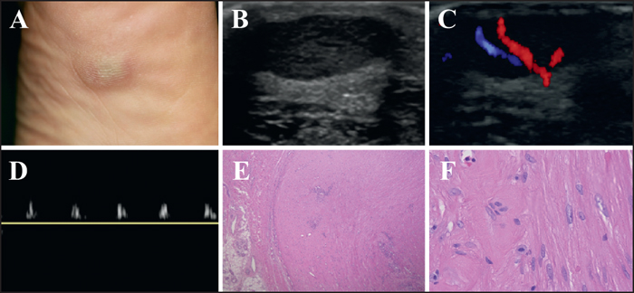
Leiomyomas are benign tumors that arise from smooth muscle. Cutaneous leiomyomas occur in three main forms: pilar leiomyoma derived from the arrector pili muscle; genital leiomyoma from smooth muscle of the scrotum, labia major or nipples; and angioleiomyoma from the media of blood vessels [1, 2]. Angioleiomyoma commonly presents as a painful solitary lesion, usually in the subcutis or, rarely, in the deep dermis [2]. Acral lesions including the fingers, toes, hands and feet are rare [2]. Here, we [...]